Türkçe
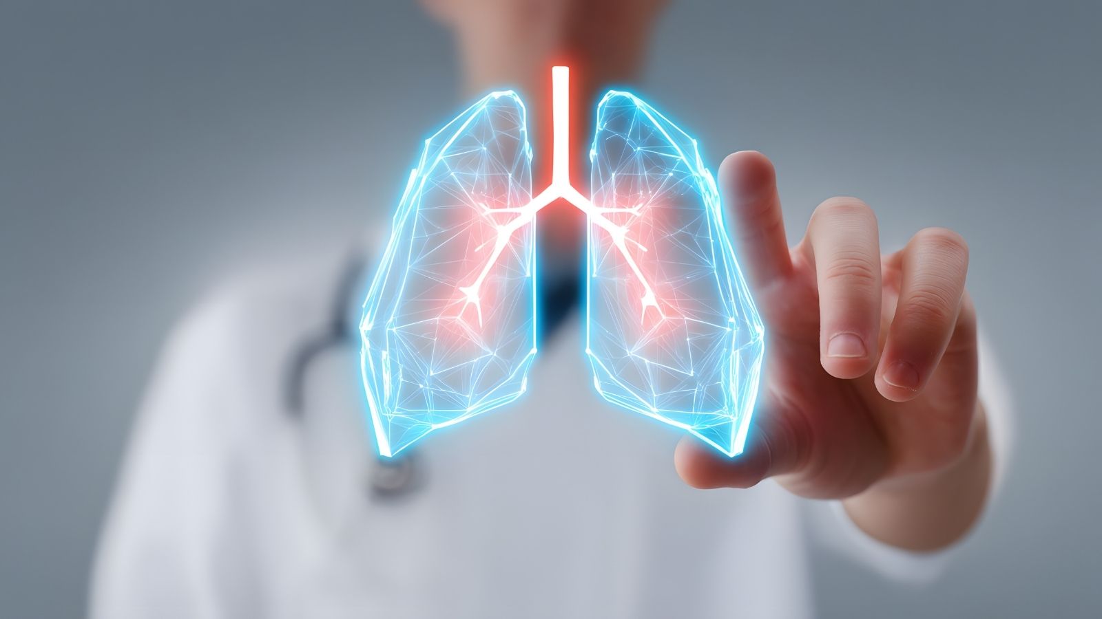
Benign lung tumors are non-cancerous cellular growths located within the chest cavity that generally carry no malignant potential and are most often detected incidentally during routine radiological examinations. Frequently reported in clinical findings as solitary pulmonary nodules, these lesions are usually the result of the body’s natural protective response to previous infections or systemic inflammatory processes. With various pathological subtypes, these structures can be effectively managed through accurate diagnostic protocols and thoracic surgical expertise without compromising respiratory health. Although symptoms are absent in most cases, modern treatment options tailored to the lesion’s location help preserve lung tissue to the greatest extent possible while safely maintaining patients’ quality of life and overall comfort.
What Do Benign Lung Tumors or Nodules Mean?
Known medically as solitary pulmonary nodules, these structures are localized areas of increased density surrounded by healthy lung tissue, typically measuring no more than three centimeters in diameter and having a round or oval shape. These lesions usually remain silent, without causing enlargement of nearby lymph nodes or fluid accumulation around the lungs. Even in completely healthy adults with no symptoms, these small lesions can be found in nearly half of screening CT scans. Approximately ninety percent of these focal cellular accumulations evaluated in thoracic surgery and pulmonology clinics are benign. In other words, they do not tend to spread to other parts of the body (metastasize) or aggressively invade surrounding tissues.
How Do Benign Lung Tumors Develop and What Causes Them?
With every breath we take, our lungs encounter thousands of microorganisms, dust particles, and foreign substances present in the air. Our immune system fights these unwanted invaders by isolating them and enclosing them within a type of cellular barrier. At the end of this healing process, a scar remains within the tissue. Just as a deep skin wound leaves behind a scar after healing, lung tissue preserves the traces of previous diseases in the form of nodules.
The main causes of these formations include:
- Tuberculosis
- Fungal infections
- Rheumatoid nodules
- Sarcoidosis
- Lung cysts
- Abscesses
Each of the conditions listed above triggers a defensive response within the lungs. For example, a previous tuberculosis infection that may have gone completely unnoticed can appear years later as a small calcified nodule on a CT scan. Likewise, inhaling fungal spores such as Aspergillus can lead to the formation of well-defined, round fungal masses (aspergilloma) within the lungs. Not only infections but also inflammatory conditions caused by the body’s own immune system, such as sarcoidosis and rheumatic diseases affecting the joints, may contribute to the development of similar benign nodules in the lungs.
How Are Benign Lung Tumors Distinguished from Malignant Masses?
When a lung lesion is detected, physicians always begin by ruling out the most serious possibility. This is because the likelihood that an incidentally discovered nodule may be malignant cannot be ignored. To make this distinction, the patient’s medical history, lifestyle, and radiological findings are evaluated with meticulous attention.
The main risk factors considered during clinical evaluation include:
- Age
- Smoking
- Asbestos exposure
- Family history
Looking more closely at these factors, advanced age and a long history of heavy smoking are among the strongest indicators raising suspicion. Occupational exposure to substances such as asbestos and a family history of lung cancer also warrant closer examination of the lesion.
In addition to these risk factors, the lesion’s appearance on computed tomography provides valuable clues regarding its nature. Benign lesions usually have smooth, well-defined borders and often contain dense calcium deposits known as calcifications. These calcifications may appear as “popcorn,” laminated, or central calcification patterns on imaging and are generally regarded as signs that the lesion has been present for a long time and has been successfully contained by the immune system. In contrast, malignant lesions tend to develop irregular, spiculated, or star-shaped margins as they invade surrounding healthy tissue. Their internal structure is typically asymmetrical and may contain thick, irregular cavities. Furthermore, the presence of small satellite nodules near the main lesion usually suggests an active or previous infectious process in that region.
How Fast Do Benign Lung Tumors Grow?
Time is one of the most reliable ways to understand the nature of a lung lesion. A single CT scan may not provide a definitive answer, which is why follow-up imaging at regular intervals is recommended. Physicians calculate how long it takes for the lesion to double in volume based on changes in its diameter. This growth rate provides valuable information about the biological behavior of the tumor.
The general classifications according to growth rate include:
- Acute
- Malignant
- Slow-growing
To explain this further, if a lesion doubles in volume within less than twenty days, the process is generally much faster than the growth rate of cancer cells. Such rapid enlargement most commonly indicates an active infection, abscess, or bleeding within the tissue. Conversely, if it takes more than four hundred days for a lesion to double in size, it is highly likely to represent stable scar tissue or an extremely slow-growing benign tumor. However, if the doubling time falls between twenty and four hundred days, the lesion is considered potentially suspicious and requires further evaluation with more advanced diagnostic tests. This mathematical assessment plays an important role in clinical decision-making and helps avoid unnecessary biopsies.
How Are Benign Lung Tumors Monitored?
Patients with low- or intermediate-risk lung nodules are not immediately referred for surgery or biopsy. Instead, scheduled CT scans are performed to monitor the lesion over time. To standardize this follow-up process and establish a common international reporting language, systems such as Lung-RADS have been developed. These systems aim to protect patients from unnecessary radiation exposure and invasive procedures.
The classification categories include:
- Category 1
- Category 2
- Category 3
- Category 4
Category 1 indicates the absence of suspicious findings and the presence of entirely benign calcified lesions, requiring no additional intervention beyond routine annual health screenings. Category 2 includes very small nodules with an extremely low clinical risk, for which annual CT surveillance is generally sufficient. Category 3 refers to nodules that are probably benign but somewhat larger in size, and patients in this group are usually advised to undergo a low-dose follow-up CT scan after six months. Category 4 and above represent lesions with suspicious features that require further diagnostic evaluation. This systematic approach ensures that each patient receives appropriate follow-up at the correct time.
What Are the Most Common Benign Lung Tumors?
Although the exact reasons why cells sometimes deviate from their normal growth patterns remain unclear, we know that benign lung tumors arise from specific tissue types. Only a very small percentage of all pulmonary neoplasms are benign, but this group includes several distinct pathological subtypes.
The most commonly encountered types include:
- Hamartomas
- Lipomas
- Leiomyomas
- Hemangiomas
Among these, hamartomas are by far the most common benign lung tumors. Typically diagnosed around the age of fifty and occurring slightly more frequently in men, these tumors consist of an abnormal mixture of the body’s own tissues, including cartilage, fat, connective tissue, and muscle cells. They are generally located in the peripheral regions of the lungs and rarely exceed a few centimeters in size. Since they usually cause no symptoms such as cough or shortness of breath, they are most often discovered incidentally and tend to grow very slowly.
Lipomas are rare tumors arising from fat cells within the airways. Leiomyomas originate from smooth muscle cells, whereas hemangiomas develop from vascular tissue. Some adenomas previously considered benign, such as carcinoid tumors, are now classified as low-grade malignant neoplasms due to advances in modern pathology. Therefore, thorough pathological examination remains critically important.
What Symptoms Do Benign Lung Tumors Located in the Airways Cause?
The location of the tumor within the lungs significantly influences the patient’s symptoms. Tumors growing within the peripheral spongy lung tissue may remain silent for years. However, tumors located within the major airways (bronchi) or directly compressing these airways may cause symptoms much earlier, even when they are benign and slow-growing.
The main symptoms may include:
- Cough
- Wheezing
- Fever
- Sputum production
- Pain
These symptoms result from a mechanical process. As an airway tumor, such as a lipoma, enlarges, it obstructs airflow through the affected bronchus. Just as air passing through a narrowed pipe produces a whistling sound, patients may hear wheezing when they breathe, often leading to confusion with asthma. As the obstruction worsens, the lungs’ natural secretions can no longer be effectively cleared by ciliated cells. The accumulated fluid behind the blockage becomes an ideal environment for bacterial growth. As a result, patients may develop recurrent episodes of pneumonia affecting the same region, accompanied by fever, severe coughing, and infected sputum. Complete obstruction eventually prevents air from reaching that portion of the lung, causing collapse (atelectasis). If this condition persists, permanent and irreversible lung damage may develop.
How Are Benign Lung Tumors Diagnosed?
No matter how detailed radiological images may be, confirming the diagnosis of a lung lesion requires obtaining tissue samples for microscopic examination. Today, thoracic surgeons and pulmonologists have advanced technologies that allow these procedures to be performed comfortably without opening the chest.
The primary diagnostic methods include:
- Bronchoscopy
- EBUS
- Biopsy
- Computed tomography
If the lesion is located near the central airways, physicians can directly examine the airways using a flexible bronchoscope equipped with a camera and light, inserted through the mouth or nose. For deeper lesions and lymph nodes located behind the airway walls, which cannot be visualized by standard bronchoscopy, a more advanced technique called EBUS (Endobronchial Ultrasonography) is used. The tip of the EBUS scope contains a miniature ultrasound probe that visualizes tissues beneath the airway surface. By viewing the exact location of the lesion and its relationship to nearby blood vessels, physicians can safely obtain tissue samples using a fine needle passed through the instrument. Procedures that previously required surgical incisions in the neck and general anesthesia can now often be completed on an outpatient basis, allowing patients to return home the same day.
If the tumor is located near the outer surface of the lung, it cannot be reached through the airways. In such cases, tissue samples are obtained by inserting a very thin needle through the chest wall under CT guidance. This procedure, known as transthoracic biopsy, is performed under local anesthesia and provides highly reliable diagnostic results. Although a small air leak may occasionally occur at the needle entry site, it is generally easily managed under medical supervision.
Which Surgical Methods Are Used to Treat Benign Lung Tumors?
If biopsy confirms that the tumor is benign, such as a hamartoma, and it is small without causing infection or airway obstruction, surgery is generally unnecessary. Instead, periodic CT scans are performed to monitor the lesion. However, if the mass continues to enlarge, causes recurrent pneumonia by obstructing the airways, steadily increases in size, or remains suspicious for malignancy despite biopsy, surgical removal becomes the safest treatment option.
The main surgical techniques include:
- Enucleation
- Wedge resection
- Segmentectomy
- Sleeve resection
The primary goal of surgery for benign lung disease is to preserve as much healthy lung tissue as possible, since the lungs are the body’s source of oxygen. Enucleation refers to removing only the tumor itself, much like extracting the core of a fruit. Wedge resection involves removing the lesion together with a very small margin of surrounding healthy tissue in a wedge-shaped fashion. When the tumor is located near the outer surface of the lung, these procedures are typically performed using Video-Assisted Thoracoscopic Surgery (VATS), a minimally invasive technique. Cameras and long, slender surgical instruments are inserted through one or several small incisions between the ribs without spreading the chest wall.
When the tumor is located deeper within the lung, wedge resection alone may result in significant air or blood leakage from the remaining tissue. Since the lungs are divided into anatomical units called segments, each supplied by its own bronchus and blood vessels, segmentectomy involves removing only the affected segment together with its dedicated airway and vascular supply. Today, these procedures can be performed using VATS or Robotic-Assisted Thoracic Surgery (RATS), in which the surgeon controls robotic instruments from a console while viewing the operative field in three dimensions.
If the tumor is located within the main bronchus, previous approaches often required removing the entire healthy lung beyond the obstruction. Today, an advanced procedure called sleeve resection is frequently performed instead. Similar to cutting out only the damaged section of a pipe and reconnecting the healthy ends, the affected portion of the airway is removed and reconstructed, preserving the patient’s healthy lung tissue.
What Can Be Expected After Surgery for Benign Lung Tumors?
The development of minimally invasive surgical techniques has made postoperative recovery considerably more comfortable than in previous decades. Since the ribs are not separated and muscle damage is minimized, patients experience significantly less pain after surgery.
Key aspects of postoperative care include:
- Drainage
- Respiratory care
- Mobilization
- Follow-up
At the end of the operation, a temporary thin chest drain (silicone tube) is inserted to remove any air or fluid that may accumulate within the chest cavity. This tube is usually removed within a few days once the lung has fully expanded and fluid drainage has ceased. Patients are encouraged to get out of bed on the first postoperative day, take short walks, and perform deep breathing exercises using respiratory training devices such as an incentive spirometer (Triflo). These simple yet effective exercises greatly contribute to restoring normal lung capacity after surgery.
Patients whose benign tumors have been completely removed are not expected to experience significant long-term health limitations. Nevertheless, because their lungs have previously developed such cellular changes and potential risk factors such as smoking may still exist, regular outpatient follow-up appointments are recommended. The purpose of this ongoing surveillance is to protect not only against recurrence of the treated condition but also to ensure the long-term health of the lungs under professional medical supervision.
Frequently Asked Questions

Prof. Dr. Alper Fındıkcıoğlu graduated from Istanbul University Faculty of Medicine and completed his thoracic surgery residency at Dr. Siyami Ersek Hospital. He began his academic career at Başkent University in 2005; he received the title of associate professor in 2014 and professor in 2022. He has extensive experience in lung cancer, chest wall diseases, and minimally invasive surgery. Since 2024, he has been continuing his professional work at Acıbadem Hospital.