Türkçe
If one lung is removed, the other healthy lung in the body expands over time and increases its volume to meet the body’s entire oxygen requirement on its own, allowing the person to continue daily life in its natural flow. After this major surgical operation known in medical language as pneumonectomy, the respiratory system restructures itself in a short time with a tremendous anatomical adaptation ability. This life-saving intervention, which is applied in diseases that threaten life or where tissue has been irreversibly damaged, does not mean being condemned to shortness of breath. Thanks to the enormous reserve respiratory capacity that the human body naturally possesses by design, patients can continue to live an active, mobile, and completely independent life.
Which Diseases Require the Removal of One Entire Lung?
The decision to completely remove one lung from the body is made after very detailed examinations and generally comes to the agenda in situations where the disease has spread too extensively to be cleaned with only a small regional operation. Malignant tumors are among the leading diseases that make this operation necessary. If the tumor is located in the exact center of the organ, has deeply progressed into the main airway, or has surrounded the large main vessels carrying blood to the lung, removing the entire lung becomes the safest way to avoid leaving diseased tissue behind. In non-cancerous conditions, situations known in the medical world as “destroyed lung,” where the tissue has irreversibly lost its structural integrity, are generally encountered. Severe infections experienced in the past may disrupt the structure of the organ and turn it into a nonfunctional source that constantly produces inflammation.
The main conditions that may require complete removal of the lung are as follows:
- Lung cancer
- Pleural mesothelioma
- Advanced tuberculosis
- Widespread bronchiectasis
- Severe fungal infections
- Serious chest trauma
Especially in conditions called destroyed lung, the diseased organ has already long abandoned its duty of supplying oxygen to the body. Not only that, but by constantly producing infection, it consumes the patient’s general energy, disrupts blood values, and tires the immune system. In such patients, removing this nonfunctioning and harmful tissue from the body, far from creating shortness of breath, allows the patient to feel much more vigorous and relieved after surgery because it removes that heavy inflammatory burden from the body. Just as a rotten fruit is removed from the basket to prevent it from spoiling the other fresh fruits, removing the destroyed organ is also essential for protecting the health of the rest of the body.
How Is It Determined That the Body Is Ready to Live with One Lung?
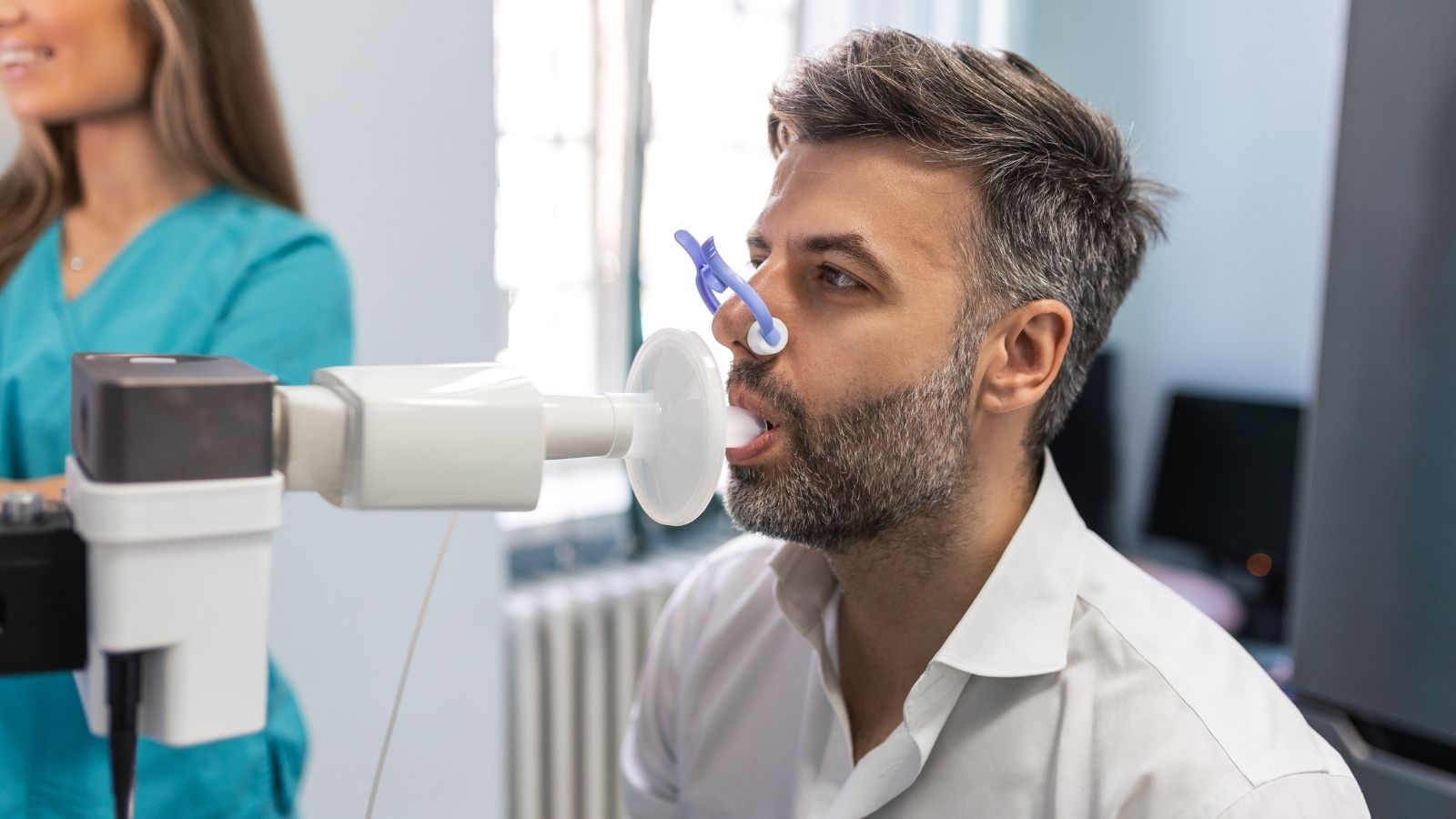
Before such an extensive surgery, the most vital preparation is calculating with mathematical certainty whether the patient can continue life safely and comfortably with a single lung. In this process, not only the condition of the diseased area but also the capacity of the healthy lung that will remain, the pumping power of the heart, and the patient’s overall physical performance are evaluated as a whole. If it is determined that the remaining tissue does not have the strength to manage the entire body, the surgical plan is changed to avoid putting the patient at risk. Very sensitive tests measuring breathing strength are applied while making this decision. The patient is asked to take a deep breath and blow into a device with all their strength. The amount of air that can be expelled within the first second is the most valuable guide in predicting the capacity of the remaining lung.
The basic measurement methods used during this evaluation stage are as follows:
- Pulmonary function tests
- Lung perfusion scintigraphy
- Stair climbing test
- Six-minute walk test
- Blood gas analysis
- Echocardiography
If the values obtained in the breathing tests are borderline, then special imaging studies showing which region of the lung is functioning and to what extent are performed for a more detailed calculation. Formulas are used to determine how many milliliters of breathing volume will remain when the diseased lung is removed. However, only lung capacity is not sufficient. Removing one lung means canceling the vessels in that region as well, and this creates a new workload on the heart. Very practical but highly reliable stair-climbing tests are applied to understand whether the heart can tolerate this burden. The number of floors the patient can climb without becoming breathless or stopping is the clearest indicator of the performance the heart will show on the operating table and during the postoperative recovery period.
Why Are Closed Surgical Methods Advantageous in Single Lung Surgery?
These surgeries, which in past years required the chest cage to be opened very widely, can now be performed extremely successfully with camera-assisted closed methods thanks to advances in technology and medical science. This modern approach called video-assisted thoracoscopic surgery is a revolutionary development that completely changes the recovery process. In open surgeries, the ribs must be separated from each other with metal instruments in order to reach the chest cavity, and this procedure may cause damage to the sensitive nerves passing beneath the ribs and lead to long-lasting postoperative pain. In the closed method, however, tiny cameras and special surgical instruments are inserted through very small incisions of a few centimeters opened between the ribs.
The main advantages provided by the closed surgical method are as follows:
- Low pain level
- Early mobilization
- Short hospital stay
- Small surgical scar
- Rapid wound healing
- Low infection risk
Thanks to the high-resolution cameras inserted inside, surgeons can view the inside of the chest cavity enlarged in the finest detail on giant screens. The main vessels connecting the lung to the heart and the airway are separated safely, bloodlessly, and quickly using highly advanced intelligent instruments similar to medical staplers. If cancer surgery is being performed, the surrounding lymph nodes carrying disease risk are also meticulously cleaned with the same closed method. Since the ribs are not forcibly separated, patients feel much less pain when they wake up after surgery. This low pain level allows the patient to turn comfortably in bed, perform deep breathing exercises without fear, and easily expel secretions inside by coughing. When all these factors come together, the patient’s return to normal life is shortened by weeks.
What Can Be Expected During the Intensive Care Process After Single Lung Surgery?
After such a major physical change, the first day when the body tries to adapt to the new condition is the most critical stage of the recovery process. The patient awakened on the operating table is immediately taken to the intensive care unit for safety purposes and for very close monitoring. Here, heartbeats, blood pressure, oxygen level in the blood, and respiratory movements are monitored second by second on giant screens. The most delicate issue in this period is adjusting the fluids the patient receives intravenously with millimetric precision. The entire blood circulation, accustomed to going to two lungs, is forced to redirect to that one healthy lung after surgery. The remaining lung suddenly shoulders the entire workload and blood pressure rises in its delicate vessels. If too much water or serum is given externally during this period, fluid may accumulate inside that single lung and breathing may become extremely difficult.
The main elements monitored to keep the patient safe during this process are as follows:
- Restricted fluid therapy
- Blood pressure control
- Heart rhythm monitoring
- Regular blood gas analysis
- Chest tube monitoring
- Oxygen support
In addition to fluid restriction, bodily fluids and air that may accumulate in the operated area inside the chest cage must be safely removed. For this purpose, the flexible and thin tube placed during surgery is connected to a special underwater system standing beside you. This system prevents you from experiencing shortness of breath by balancing the internal pressure sensitively. At the same time, temporary accelerations or irregularities in heart rhythm may sometimes occur as a reaction of the body to this sudden volume change. This is an extremely expected condition and the heart rhythm is easily returned to its former calm state with medication treatments immediately applied in the intensive care environment. All this meticulous monitoring forms the solid foundations laid for the body to safely adapt to its new anatomy.
If One Lung Is Removed, How Does the Body Adapt to This Empty Space?
When one lung is completely removed from the body, many people wonder how the large space created inside will be filled and how the body will maintain its balance. The anatomical adaptation ability of the human body comes into play exactly at this point and works almost like a marvel of engineering. The right lung consists of three sections, while the left lung consists of two sections. Depending on which side is removed, the remaining space inside is gradually filled by the body through its own natural mechanisms. During the first months after surgery, this space is slowly filled with a sterile and clean fluid naturally produced by the body. As time progresses, this fluid solidifies into a gelatinous structure and eventually transforms into what we call fibrotic connective tissue, permanently filling and stabilizing the area.
The physical changes the body exhibits while adapting to this space are as follows:
- Expansion of the remaining lung
- Filling of the chest cavity with fluid
- Hardening of tissues by solidification
- Upward pulling of the diaphragm muscle
- Slight shifting of the heart from the midline
- Narrowing of the rib spaces
Not only is the space filled, but the healthy lung continuing to function alone on the opposite side also shows incredible performance. Realizing this new and great responsibility placed upon it, the single lung gradually stretches toward the midline, pushes its limits, and physically increases its capacity. Millions of tiny air sacs inside the lung open much wider than before and maximize their capacity to capture oxygen in the air. This tremendous expansion and tissue growth process is completed in approximately six months to one year. Once the process is completed, you become almost unable to feel any difference from your previous condition while resting or performing ordinary daily tasks. The body has reestablished its tremendous balance and completely learned to live with one lung.
Which Breathing Exercises Should Be Performed to Increase Single Lung Capacity?
The success of surgery is not a process completed on the operating table; the greatest key to recovery is the investment the patient makes in their own body after surgery. The secret to transitioning to a highly efficient and quality life with one lung is to work that single lung regularly and expand its capacity. These exercises, which begin in the hospital during those first days under the guidance of respiratory physiotherapists, should also become part of your daily routine at home after discharge. The exercises taught aim to transfer the work of breathing from the tiring burden of the chest muscles to the diaphragm, a much stronger and more durable muscle. Thus, each breath is taken much more economically and with far less energy expenditure.
The exercises that should be performed regularly every day at home are as follows:
- Diaphragmatic breathing exercise
- Pursed-lip breathing
- Breathing with a triflo device
- Shoulder rotation movements
- Light-paced walking
- Cough support techniques
Especially diaphragmatic breathing is very effective. This style of breathing, performed by pushing your abdomen outward while inhaling instead of inflating your chest, allows you to actively use that large diaphragm muscle located below. This guarantees that even the lowest and most prone-to-collapse parts of the lung are filled with perfectly clean air. The pursed-lip technique you apply when you feel short of breath is a lifesaver. When you inhale deeply through your nose and slowly exhale while pursing your lips as if whistling, you increase the pressure inside the airways. This increase in internal pressure prevents the tired airways from closing prematurely, facilitates the complete expulsion of trapped stale air inside, and creates room for your lung to take in more fresh air in the next breath.
How Should One Eat to Stay Healthy with a Single Lung?
After a life-changing surgery, the body must both heal its wounds and keep the remaining respiratory muscles very strong. This is only possible with a properly planned, quality nutritional routine. The biggest mistake made in postoperative nutrition is consuming too much and very heavy meals at once with the thought of rapid recovery. The single lung needs space to expand in order to function healthily. If you overfill your stomach in a single meal, the swollen stomach pushes upward against the diaphragm muscle. The upward-pushed diaphragm then prevents the lung from expanding comfortably, causing you to feel mechanical shortness of breath. Therefore, the nutritional order should be completely reorganized into small but frequent meals.
Foods that support respiratory muscles and accelerate tissue repair are as follows:
- Eggs
- Lean red meat
- Grilled fish
- Boiled chicken
- Fresh green leafy vegetables
- Citrus fruits containing vitamin C
Protein, in particular, is the indispensable building block of this process. Breathing is a muscular activity, and sufficient amounts of quality protein must be consumed every day so that the chest muscles serving the single lung do not waste away over time and maintain their endurance. Instead of heavy cooking methods such as frying, baking or boiling should be preferred in meat consumption. At the same time, fruits and vegetables rich in vitamins C and E, which support cellular repair and clean oxidative damage in the body, should be placed at the center of nutrition. Staying away from carbonated beverages that cause bloating, legumes that produce excessive gas, and pastries will help you breathe much more comfortably throughout the day because it eliminates the pressure the stomach exerts on the lung.
What Should Be Considered in Lifestyle After Single Lung Surgery?
When you leave the hospital and return home, you must now adopt a brand-new and much more conscious lifestyle. Maximum care should be taken to protect that single lung so it can carry the burden of the entire body smoothly. The sharpest and most indisputable rule of this new life is airway cleanliness. Smoking and all tobacco products must be completely removed from life never to be considered again. It is not enough that you yourself do not smoke; even being in closed, airless, smoke-filled environments where someone else is smoking is one of the greatest harms that can be done to your remaining lung. The damage caused by passive smoking is far more devastating for a person with one lung than what a healthy individual could tolerate.
The basic precautions you should take to protect your lung in daily life are as follows:
- Staying away from smoking environments
- Getting an annual flu vaccine
- Receiving the pneumonia vaccine
- Wearing a mask in crowded environments
- Taking regular walks in forests
- Cleaning the air in the house every day
You should be one level more alert than normal against infections. Even a simple cold may turn into fatigue more quickly in a single-lung system. Therefore, getting your seasonal vaccines completely before winter approaches and definitely using a protective mask when entering public transportation or crowded closed spaces is a very smart defense strategy. Regular walks in parks or forested areas with as much clean, oxygen-rich air as possible, without tiring yourself, will maximize the oxygen-carrying capacity of the lung.

Prof. Dr. Alper Fındıkcıoğlu graduated from Istanbul University Faculty of Medicine and completed his thoracic surgery residency at Dr. Siyami Ersek Hospital. He began his academic career at Başkent University in 2005; he received the title of associate professor in 2014 and professor in 2022. He has extensive experience in lung cancer, chest wall diseases, and minimally invasive surgery. Since 2024, he has been continuing his professional work at Acıbadem Hospital.