Türkçe
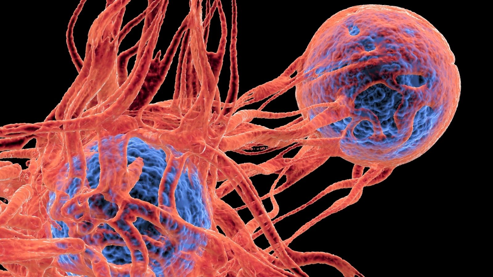
Adenocarcinoma is a malignant type of cancer that develops from glandular tissue and originates from mucus- or secretion-producing cells. It most commonly occurs in organs such as the lungs, stomach, pancreas, colon, and prostate. The disease progresses through uncontrolled cell proliferation and, if not diagnosed early, can spread to surrounding tissues and distant organs.
The symptoms of adenocarcinoma vary depending on the affected organ. When it develops in the lungs, it may cause a persistent cough, shortness of breath, and chest pain, whereas intestinal adenocarcinoma may present with changes in bowel habits, abdominal pain, and weight loss. Fatigue and loss of appetite are also common in advanced stages.
The diagnosis of adenocarcinoma is established through imaging methods and pathological examination. Computed tomography, magnetic resonance imaging, and endoscopic evaluations are used to determine the location and extent of the tumor. A definitive diagnosis is usually confirmed by microscopic examination of a tissue sample obtained through biopsy.
Treatment methods for adenocarcinoma are planned according to the stage of the disease, the organ involved, and the patient’s overall health status. Surgery, chemotherapy, radiotherapy, and targeted therapies are the most commonly used treatment approaches. In cases diagnosed at an early stage, treatment success and survival rates can increase significantly.
| What You Need to Know | Information |
| Definition of the Disease | Adenocarcinoma is a malignant (cancerous) tumor that develops from glandular cells and typically affects mucus-producing cells. It can occur in various organs, including the lungs, colon, stomach, pancreas, prostate, breast, and others. |
| Common Sites | It is most commonly found in the lungs, colon (large intestine), stomach, pancreas, prostate, and breast tissue. |
| Risk Factors | Genetic predisposition, smoking and alcohol use, obesity, advanced age, unhealthy dietary habits, chronic inflammation, and exposure to environmental toxins. |
| Symptoms (General) | – Unexplained weight loss – Weakness and fatigue – Persistent pain – Bloody stool or sputum – Persistent cough or hoarseness – Abdominal pain, loss of appetite – Abdominal swelling or a palpable mass – Jaundice (in pancreatic adenocarcinoma) |
| Symptoms (Organ-Specific) | – Lung: Persistent cough, bloody sputum, shortness of breath – Colon: Changes in bowel habits, blood in the stool – Stomach: Stomach pain, early satiety, weight loss – Pancreas: Back pain, jaundice, weight loss – Prostate: Difficulty urinating, pelvic pain – Breast: Lump, nipple discharge, or nipple retraction |
| Diagnostic Methods | – Physical examination – Blood tests – Biopsy – Imaging methods: CT (Computed Tomography), MRI, PET scans, endoscopy, colonoscopy – Tumor markers (especially those specific to certain organs) |
| Treatment Methods | – Surgical intervention (removal of the tumor in early-stage disease) – Chemotherapy – Radiotherapy – Targeted therapy – Immunotherapy (particularly for certain lung and colon adenocarcinomas) – Palliative care (to manage symptoms in advanced cases) |
| Prognosis (Disease Course) | It varies depending on the affected organ, the stage at diagnosis, and the response to treatment. Early diagnosis significantly improves treatment success. |
| Follow-up and Monitoring | Regular screening and follow-up tests are important to monitor for recurrence after treatment. Periodic evaluations are performed following treatment. |
| Prevention | Maintaining a healthy diet, avoiding smoking and alcohol, exercising regularly, participating in screening programs (especially for high-risk individuals), and minimizing environmental risk exposure. |
What Is Lung Adenocarcinoma?
The human respiratory system can be imagined as a giant upside-down tree. The main trachea forms the trunk, while inside the lungs it branches into increasingly smaller airways that ultimately end in tiny air sacs resembling leaves. Lung adenocarcinoma originates from the secretory cells lining these microscopic air sacs.
These cells constantly function to trap inhaled particles and prevent the airways from drying out. However, over time, the accumulation of various external factors or internal genetic abnormalities can damage the DNA of these beneficial cells.
Once the cell’s regulatory mechanisms fail, it no longer recognizes the rule that it should remain in its original location. While normal cells are eliminated by the body after completing their lifespan, these mutated cells continue dividing as though they have become immortal. They initially form microscopic clusters and eventually develop into structures identified on imaging studies as nodules or masses. The greatest danger of these tumors is not only their growth but also their ability to invade nearby blood vessels and lymphatic channels.
What Are the Main Risk Factors for Developing Lung Adenocarcinoma?
When people think of lung cancer, they often associate it with individuals who have smoked heavily for many years. The thousands of toxic and carcinogenic chemicals found in tobacco smoke directly damage the genetic material of lung cells. However, adenocarcinoma presents a unique epidemiological pattern that differs from this traditional understanding. In recent years, worldwide health data have demonstrated a significant increase in this disease among people who have never smoked, particularly women.
Beyond tobacco products, modern environmental factors also play a substantial role in cellular damage. Radon, a colorless and odorless gas that accumulates indoors and seeps from the ground, represents a silent hazard. Likewise, inhalation of asbestos fibers found in older insulation materials causes permanent damage to the lungs and pleural tissue.
Risk factors include:
- Tobacco use
- Secondhand smoke exposure
- Radon gas
- Asbestos exposure
- Air pollution
- Advanced age
- Genetic predisposition
What Symptoms Are Experienced in Early-Stage Lung Adenocarcinoma?
The anatomical structure of the lungs contains very few nerve endings responsible for transmitting pain signals to the brain. Pain-sensitive nerves are primarily located in the pleural membrane surrounding the lungs and within the chest wall. Since adenocarcinomas characteristically develop in the deep outer (peripheral) regions of the lungs, they usually do not compress surrounding structures or cause pain during the early stages. Consequently, the uncontrolled cellular growth progresses silently without producing noticeable symptoms.
Most patients continue their daily lives believing they are completely healthy. Tumors at this stage are often discovered incidentally during chest X-rays or CT scans performed for unrelated reasons, such as pre-employment examinations, routine preoperative evaluations, or investigations for common respiratory infections. This situation, referred to medically as an “incidental pulmonary nodule,” actually represents the most treatable and favorable stage of the disease. This silent progression clearly demonstrates the life-saving importance of regular health screenings for individuals at increased risk.
What Symptoms Does Advanced Lung Adenocarcinoma Cause?
If the disease is not detected and treated in time, the tumor gradually enlarges. As it grows, it may narrow the airways, involve the sensitive pleural membranes surrounding the lungs, or directly invade the muscles and bones of the chest wall.
Irritation of the airways by the growing tumor often triggers the body’s natural reflex to expel a foreign substance, resulting in a persistent cough.
Certain tumors located in the upper portions of the lungs may also lie close to the major nerve networks supplying the neck and arms. When these nerves are compressed, patients may experience severe and persistent pain radiating from the shoulder or back down the arm to the fingertips rather than pain originating directly from the chest. As the disease progresses, the tumor increasingly consumes the body’s energy reserves, leading to metabolic imbalance.
Symptoms of advanced disease include:
- Persistent cough
- Shortness of breath
- Chest pain
- Back pain
- Shoulder pain
- Bloody sputum
- Hoarseness
- Difficulty swallowing
- Loss of appetite
- Weight loss
- Chronic fatigue
Which Imaging Techniques Are Used When Lung Adenocarcinoma Is Suspected?
Radiological imaging plays a central role in the diagnostic process initiated by symptoms or incidental findings. Although a standard chest X-ray may reveal large masses within the chest, it is insufficient for identifying the detailed characteristics of adenocarcinoma or detecting very small nodules. High-resolution Computed Tomography (CT) is the primary imaging modality. CT scans evaluate the lungs in millimeter-thick slices, clearly revealing the size of the suspicious lesion, whether its borders are smooth or spiculated, and its relationship with surrounding blood vessels.
If detailed evaluation of the tumor’s proximity to the chest wall, spine, or nerves is required, Magnetic Resonance Imaging (MRI) is preferred. Another essential imaging study is Positron Emission Tomography combined with CT (PET/CT). Because cancer cells consume significantly more energy than normal cells, they rapidly absorb a harmless low-dose radioactive glucose compound administered before the scan. PET imaging measures the metabolic activity of the lung tumor while simultaneously determining whether the disease has spread to lymph nodes or distant organs.
How Is a Tissue Sample Obtained to Confirm the Diagnosis of Lung Adenocarcinoma?
Although imaging studies provide strong evidence suggesting that a mass may be cancerous, a definitive diagnosis can only be established by examining a tissue sample under a microscope. This procedure is known as a biopsy. The location of the tumor within the lung determines which biopsy technique is most appropriate.
If the tumor is located centrally near the main airways, bronchoscopy is usually performed. During this procedure, a thin flexible tube equipped with a camera and light source is passed through the airways, allowing direct visualization and sampling of the suspicious tissue. However, because adenocarcinomas are often situated near the outer surface of the lungs, the bronchoscope may not reach these peripheral lesions. In such cases, a transthoracic needle biopsy guided by CT imaging is a more common and successful approach. Performed under local anesthesia, this safe procedure typically allows patients to return home the same day.
How Does Staging of Lung Adenocarcinoma Affect Surgical Planning?
Once adenocarcinoma has been confirmed by biopsy, determining the stage of the disease becomes the next critical step. Staging is based on the size of the tumor, its extent within the lung, and whether it has spread to lymph nodes or distant organs. Modern pathology also classifies adenocarcinoma according to detailed microscopic growth patterns.
For example, patients whose tumors are very small and demonstrate only minimal invasion into surrounding healthy tissue are particularly fortunate. At this early stage, the disease is considered localized, and treatment success approaches nearly one hundred percent. However, when aggressive growth patterns such as micropapillary or solid subtypes are identified, the likelihood of vascular invasion increases. This finding often necessitates more extensive surgery and the addition of postoperative adjuvant therapies.
How Is Respiratory Capacity Calculated Before Lung Adenocarcinoma Surgery?
Determining whether a patient is suitable for surgery requires evaluation not only of tumor resectability but also of whether sufficient lung function will remain afterward. Removing part or all of a lung directly affects the body’s ability to oxygenate the blood. Therefore, every patient undergoes a detailed Pulmonary Function Test (PFT) before surgery.
The most important measurement is the forced expiratory volume in one second (FEV1), which represents the amount of air the patient can forcefully exhale during the first second after taking a deep breath. This value reflects the patient’s current lung function. To estimate postoperative lung function, physicians calculate the predicted postoperative FEV1 (ppoFEV1) using the following standard formula:
\text{ppoFEV1} = \text{Preoperative FEV1} \times (1 – \frac{\text{Number of Segments to Be Removed}}{\text{Total Number of Segments}})
If the calculated value falls below accepted safety thresholds, the patient may be at risk of prolonged dependence on mechanical ventilation after surgery. In such cases, the surgical plan is reassessed, cardiac function is evaluated with echocardiography, and arterial blood gas analysis is performed to determine the safest treatment strategy.
Why Are Lymph Nodes So Important in the Treatment of Lung Adenocarcinoma?
The human body contains an extensive lymphatic network that collects excess tissue fluid and serves as an important component of the immune system. When lung cancer cells begin to spread, they typically enter the lymphatic vessels and first reach the mediastinal lymph nodes located around the heart and trachea.
The presence or absence of tumor cells within these lymph nodes fundamentally changes the treatment strategy. If the disease remains confined to the lung, surgery can often be performed immediately. However, if the mediastinal lymph nodes are involved, systemic therapy is generally recommended first to reduce tumor burden and eliminate microscopic disease. Endobronchial ultrasound-guided transbronchial needle aspiration (EBUS) is currently one of the most reliable and widely used methods for evaluating these lymph nodes.
How Is Lobectomy, the Gold Standard Surgical Treatment for Lung Adenocarcinoma, Performed?
The lungs are not single solid organs. The right lung consists of three lobes, while the left lung contains two because of the space occupied by the heart. Each lobe has its own blood vessels and airway branches.
An anatomical lobectomy involves removing the entire lobe containing the tumor together with its supplying blood vessels and bronchus as a single unit. Because adenocarcinomas frequently develop near the edges of a lobe, removing the entire lobe ensures a wide margin of healthy tissue around the tumor, minimizing the risk of local recurrence. During surgery, specialized stapling devices simultaneously divide and seal blood vessels and tissues, allowing the procedure to be completed safely.
What Lung-Preserving Surgical Techniques Are Used for Limited Lung Adenocarcinoma?
Removal of an entire lung lobe is not necessary or feasible for every patient. If pulmonary function is insufficient to tolerate a lobectomy or if the tumor is very small and detected at an early stage, more limited lung-preserving procedures may be selected.
Segmentectomy involves removing only the specific anatomical segment containing the tumor while preserving the remainder of the lobe. This approach provides excellent preservation of healthy lung tissue but requires a high level of surgical expertise. Wedge resection involves removing a wedge-shaped portion of lung tissue surrounding the tumor without following anatomical segmental boundaries. It is most commonly used for diagnostic purposes or in patients with very limited pulmonary reserve.
How Is Lung Adenocarcinoma Surgery Performed Using Minimally Invasive VATS?
In the past, lung surgery required large chest incisions and forceful separation of the ribs using metal retractors, resulting in considerable postoperative pain involving both bone and muscle.
Today, Video-Assisted Thoracoscopic Surgery (VATS) has transformed this experience.
With VATS, the ribs are neither cut nor forcibly spread apart. Instead, high-definition cameras and long, slender surgical instruments are inserted through one or several small incisions in the chest wall. Rather than looking directly into the chest cavity, the surgeon performs the operation while viewing magnified images on large monitors. Using the most advanced “uniportal” technique, even an entire lobectomy can be completed through a single incision measuring only 3–4 centimeters.
The main advantages of VATS include:
- Less pain
- Smaller surgical scars
- Lower risk of infection
- Less bleeding
- Shorter hospital stay
- Earlier discharge
- Faster return to daily life
- Better preservation of the immune system
How Do Robotic Systems Improve Lung Adenocarcinoma Surgery?
Robotic surgery represents the most advanced form of minimally invasive surgery. Contrary to common belief, the robot does not perform the operation independently. The surgeon maintains complete control while operating from a specialized console within the operating room.
Robotic technology provides a high-definition three-dimensional view of the surgical field magnified up to fifteen times, offering exceptional depth perception. Natural hand tremors are completely filtered by the robotic system. The robotic instruments possess a range of motion exceeding that of the human wrist, allowing precise movements in every direction. This capability is especially valuable when operating in difficult-to-reach areas behind the heart and major blood vessels, enabling delicate tissue dissection and precise suturing with outstanding safety.
Why Must Lymph Nodes Be Removed During Lung Adenocarcinoma Surgery?
Regardless of the surgical technique used, removing the lung tumor is only one component of successful treatment. The greatest determinant of long-term survival is systematic removal of the lymph nodes within the chest. During systematic lymph node dissection, predefined lymph node stations surrounding the tumor and within the mediastinum are removed as a single specimen.
This procedure eliminates microscopic cancer cells that cannot be seen with the naked eye. After surgery, each lymph node is examined individually by pathologists to determine whether cancer cells are present. These findings establish the definitive stage of the disease and provide the strongest scientific basis for deciding whether additional postoperative treatments are necessary.
Which Drug Therapies Are Used During Recovery From Lung Adenocarcinoma?
Cancer treatment requires a multidisciplinary approach and extends beyond surgery alone. By analyzing the tumor’s genetic profile, physicians can tailor medical treatments to each patient’s specific disease characteristics. In some cases, even when the tumor appears surgically removable, chemotherapy or immunotherapy may be administered first to reduce tumor size before surgery. This approach, known as neoadjuvant therapy, aims to maximize the likelihood of successful surgical treatment.
If postoperative pathology reveals lymph node involvement or a tumor exceeding a certain size, adjuvant therapies are initiated to eliminate microscopic cancer cells that may remain elsewhere in the body. When specific genetic mutations are identified within adenocarcinoma cells, targeted oral therapies—often referred to as “smart drugs”—can substantially reduce the risk of recurrence.
How Can the Hospital Discharge Process Be Accelerated After Lung Adenocarcinoma Surgery?
Following successful surgery, enhanced recovery protocols are implemented to help patients regain their health and quality of life as quickly as possible. Pneumonia is one of the most concerning complications after lung surgery. Effective coughing is essential to clear accumulated secretions from the lungs, but pain may prevent patients from coughing adequately. Therefore, epidural catheters or regional nerve blocks are commonly used to provide effective pain control.
Patients are encouraged to get out of bed and begin walking on the evening of surgery or, at the latest, the following morning. Early mobilization promotes lung expansion while reducing the risk of dangerous blood clots in the legs. Under the supervision of respiratory physiotherapists, balloon inflation exercises and specialized breathing devices gradually restore lung capacity.
The main components of an enhanced recovery protocol include:
- Effective pain control
- Early ambulation
- Breathing exercises
- Early nutrition
- Early removal of catheters
- Muscle-strengthening exercises
How Should Follow-up Be Conducted After Lung Adenocarcinoma Treatment?
Once treatment has been completed and the patient returns home, a new phase of care begins: surveillance. If lung cancer recurs, it most commonly does so within the first two years after treatment. Consequently, follow-up is particularly intensive during this critical period.
For the first two years after treatment, patients undergo physical examinations every three months, accompanied by regular low-dose CT scans to monitor their lungs. Between the second and fifth years, follow-up intervals are extended to every six months as the risk decreases. Although patients are generally considered clinically cured after five years, lifelong annual health screening remains strongly recommended because of the underlying susceptibility of lung tissue.
Frequently Asked Questions

Prof. Dr. Alper Fındıkcıoğlu graduated from Istanbul University Faculty of Medicine and completed his thoracic surgery residency at Dr. Siyami Ersek Hospital. He began his academic career at Başkent University in 2005; he received the title of associate professor in 2014 and professor in 2022. He has extensive experience in lung cancer, chest wall diseases, and minimally invasive surgery. Since 2024, he has been continuing his professional work at Acıbadem Hospital.