Türkçe
Lung biopsy is an invasive medical procedure performed by obtaining a tissue sample from the lung for pathological examination. This method is used to establish a definitive diagnosis of lung nodules, masses, infections, and interstitial lung diseases, contributing to the development of an appropriate treatment plan.
The procedure for performing a lung biopsy depends on the patient’s clinical condition and may be carried out using bronchoscopic, needle aspiration, or surgical techniques. Image-guided needle biopsy is minimally invasive. When a more comprehensive tissue sample is required, video-assisted thoracoscopic surgery may be preferred.
Regarding the question of when a lung biopsy is indicated, lesions suspicious for malignancy, unexplained pulmonary infiltrates, and treatment-resistant infections are among the primary indications. It also plays a significant role in confirming the diagnosis of diseases such as sarcoidosis and pulmonary fibrosis.
The risks and complications of a lung biopsy include pneumothorax, bleeding, and infection. After the procedure, patients are monitored for vital signs and respiratory function. Prompt management of potential early complications is essential for ensuring a safe recovery process.
In Which Situations Is a Lung Biopsy Necessary?
In medical practice, a lung biopsy is generally considered when an abnormal structure is detected within the lung tissue during a chest X-ray performed as part of a routine health screening or a computed tomography (CT) scan requested for a more detailed evaluation. These findings are commonly referred to by the public as spots, nodules, or masses. However, not every spot identified on imaging immediately leads to a biopsy. In many cases, when such a suspicious finding is detected, antibiotic treatment is first administered for a certain period to rule out the possibility that the underlying cause is a simple pneumonia or similar infection, followed by repeat imaging. If the lesion continues to grow despite treatment, develops a more irregular appearance, or has characteristics highly suggestive of malignancy from the outset, a biopsy becomes unavoidable to determine the exact cellular structure. Particularly in serious cases where lung cancer is suspected, obtaining a tissue diagnosis is a medical necessity to identify the exact cancer type and stage, as well as to determine the most appropriate treatment.
Does Every Spot in the Lung Require a Lung Biopsy?
The answer is definitely no, and this is reassuring information for the general public. The vast majority of lesions or small nodules incidentally detected in the lungs through imaging are completely benign and pose no danger. Since the lungs are constantly exposed to the outside world, filtering numerous particles and microorganisms inhaled with the air we breathe, previous unnoticed illnesses may leave permanent marks in the lungs. Because it is not always possible to determine whether a nodule is dangerous or simply a harmless scar based solely on imaging, cellular examination may be necessary. This procedure enables early diagnosis if a serious disease is present while eliminating unnecessary concerns if the lesion is benign.
Some harmless conditions that may appear as lung spots include:
- Previous infections
- Tuberculosis scars
- Fungal diseases
- Rheumatologic involvement
- Benign cysts
- Inflammatory accumulations
Which Criteria Are Considered When Deciding on a Lung Biopsy?
The decision to perform a biopsy is made after carefully analyzing multiple factors, including the patient’s demographic characteristics, medical history, and the radiological morphology of the detected nodule. Age is one of the strongest determining factors in this process. According to medical and statistical data, pulmonary lesions detected in individuals younger than forty years of age are much less likely to be malignant. In this age group, periodic radiological follow-up is generally preferred instead of immediately performing an invasive procedure. However, as age increases, especially in individuals aged sixty years and older, the likelihood that newly detected lesions are malignant rises significantly. In this group, obtaining a definitive tissue diagnosis through biopsy becomes a much more rational and necessary medical approach rather than simply waiting.
The size of the nodule also plays a critical role in decision-making. Nodules smaller than five millimeters generally have a malignancy risk of less than one percent. Since such tiny structures are difficult to target with biopsy and carry minimal risk, monitoring with CT scans every six months or annually is usually considered sufficient. In contrast, lesions exceeding twenty millimeters carry a much higher risk and generally require prompt intervention and tissue sampling.
The main factors directly influencing the biopsy decision include:
- Patient’s age
- Nodule size
- Nodule shape
- Growth rate
- Smoking history
- Family history
What Is the Role of PET-CT Before a Lung Biopsy?
Computed tomography is an excellent standard tool for accurately identifying the anatomical location of the area from which tissue will be sampled. However, CT only demonstrates the size and shape of a structure and cannot reveal what is happening inside it or how active its cells are. Positron Emission Tomography-Computed Tomography (PET-CT) is considered the gold standard for evaluating metabolic activity at the cellular level. During this advanced imaging examination, the patient receives a very low-dose radioactive glucose molecule through a vein. Since malignant cancer cells divide rapidly and uncontrollably, they require significantly more energy and therefore absorb much more glucose than normal healthy cells.
The scanner measures how intensely the suspicious lung lesion absorbs this glucose using a standardized value. If uptake exceeds established thresholds, the likelihood of malignancy increases considerably, making biopsy clearly indicated. Another critical role of PET-CT is guiding the physician to the most appropriate biopsy site. Large tumors may contain areas of dead tissue, while PET-CT highlights the most metabolically active region, preventing sampling from non-diagnostic areas and reducing delays in diagnosis.
In Which Situations Is Nodule Surveillance Preferred Instead of Lung Biopsy?
If CT scans, PET imaging, and other evaluations indicate that a pulmonary nodule is not suspicious enough to warrant immediate intervention, a carefully controlled observation period is initiated. This process does not mean abandoning the patient; rather, it involves close radiological monitoring to detect any changes in size, cellular activity, or morphology. Completely solid nodules are typically monitored at regular intervals for two years. If no change in size or appearance occurs during this period, they are generally considered benign and follow-up intervals may be extended.
However, subsolid nodules, often referred to as ground-glass nodules, behave differently. Since they may grow much more slowly, two years of follow-up may not be sufficient. Therefore, surveillance is generally extended to at least three years. Any increase in the solid component or changes in density during follow-up are considered indications for biopsy. Nodules containing calcification are generally benign and usually require no further follow-up unless they exhibit suspicious characteristics.
How Is a Needle Lung Biopsy Performed?
Needle biopsy, medically known as percutaneous biopsy, is commonly used for lesions located near the outer portions of the lungs, close to the chest wall and ribs rather than centrally. The procedure is generally performed in the interventional radiology unit of a fully equipped hospital under real-time CT or ultrasound guidance. It requires advanced technology and highly precise planning. While the patient lies comfortably on the CT table, the scanner identifies the exact three-dimensional coordinates of the lesion within the chest.
The skin over the biopsy site is completely numbed with a powerful local anesthetic, preventing the patient from feeling the needle. A long, thin specialized needle is then carefully advanced between the ribs directly into the lung lesion. Once the needle reaches the target, cellular material or tiny tissue fragments are aspirated into a syringe using negative pressure, completing the procedure. This technique is particularly valuable for diagnosing peripheral lung lesions that cannot be reached using bronchoscopy.
What Are the Differences Between Fine Needle and Core Needle Lung Biopsy?
The thickness and design of the biopsy needle directly affect the quality of the tissue sample and, consequently, the accuracy of the pathological diagnosis. Fine needle aspiration biopsy uses extremely thin needles similar to those used for blood sampling. Cells are aspirated from the lesion into a syringe. This technique is highly effective and safe for detecting malignant cells. However, because the cells are removed individually or in small clusters, the original tissue architecture is not preserved.
Core needle biopsy, commonly referred to as Tru-cut biopsy, is different. These needles contain a spring-loaded cutting mechanism that removes a small cylindrical core of tissue while preserving its structural integrity. The greatest advantage of this method is that it allows the pathologist to evaluate not only individual cells but also the overall tissue architecture, blood vessels, and surrounding structures. Core needle biopsy is therefore often preferred for diagnosing certain lymphomas and distinguishing various benign diseases.
When Is Surgical Lung Biopsy Necessary?
Although needle biopsies are advanced and minimally invasive, they do not always provide a definitive diagnosis. Sometimes a lesion is located in an anatomically hazardous region, such as immediately adjacent to the heart or major pulmonary vessels, making needle biopsy unsafe. In other cases, the lesion may simply be too small to target accurately, or the patient’s lung anatomy may not be suitable for needle biopsy. Furthermore, previous needle biopsies may have yielded insufficient tissue for a definitive pathological diagnosis.
In these situations, or when the healthcare team intends not only to diagnose but also to completely remove the lesion during the same procedure, surgical techniques performed by thoracic surgeons become necessary. Modern minimally invasive thoracic surgical approaches offer the highest diagnostic accuracy, approaching nearly one hundred percent while simultaneously providing definitive treatment when appropriate.
What Benefits Does Minimally Invasive Surgical Lung Biopsy Offer Patients?
Video-Assisted Thoracoscopic Surgery (VATS) is a revolutionary advancement in thoracic surgery that has dramatically improved patient comfort. In the past, obtaining even a small lung tissue sample required opening the chest widely and spreading the ribs with metal instruments. Today, this is no longer necessary. Under general anesthesia, surgeons make only one to three small incisions between the ribs, each measuring approximately one to three centimeters. High-definition cameras and specialized surgical instruments are inserted through these openings, allowing the surgeon to visualize the chest cavity on large monitors with exceptional clarity.
This minimally invasive approach offers tremendous advantages throughout the patient’s recovery. Because the extensive tissue damage associated with traditional open surgery is avoided, healing is significantly faster.
The main advantages of this technique include:
- Definitive diagnosis
- Less pain
- Faster recovery
- Cosmetic benefit
- Simultaneous treatment
- Lower infection risk
Why Is Lung Biopsy Performed for the Pleura?
Not only the lungs themselves but also the thin double-layered membrane surrounding them, known as the pleura, can become affected by disease. In healthy individuals, only a small amount of lubricating fluid exists between these layers to reduce friction during breathing. However, conditions such as lung cancer spreading to the pleura, asbestos-related pleural malignancies, or severe untreated tuberculosis may damage these membranes. As a result, excessive fluid accumulates between them, compressing the lung and causing shortness of breath.
Although fluid can initially be sampled with a needle, it may not contain enough cells for a definitive diagnosis. In such cases, obtaining a direct biopsy from the pleural tissue becomes necessary. During minimally invasive surgery, the surgeon examines the entire pleural surface using a camera and directly samples abnormal thickened or irregular areas. In addition, medications can be applied to permanently adhere the pleural layers together, preventing fluid from recurring and relieving symptoms.
What Is Mediastinal Staging Before a Lung Biopsy?
If lung cancer is strongly suspected or already confirmed by needle biopsy, the status of the mediastinal lymph nodes becomes the most critical factor in determining treatment. The mediastinum is the central compartment between the lungs containing the heart and major blood vessels. Lymph nodes act as filters, and cancer cells often spread there first. If cancer has already reached these lymph nodes, systemic treatment such as chemotherapy is generally more appropriate than immediate surgery.
To determine whether these lymph nodes contain cancer cells, a specialized surgical procedure known as mediastinoscopy is performed. Thoracic surgeons make a small horizontal incision approximately two centimeters long just above the breastbone and insert a lighted camera in front of the trachea. Without opening the chest, suspicious lymph nodes can be directly visualized and biopsied. Because this area contains major blood vessels and nerves controlling the vocal cords, extensive anatomical knowledge and surgical expertise are essential for patient safety.
What Should Patients Pay Attention to Before a Lung Biopsy?
Whether the biopsy is performed under local anesthesia in the interventional radiology department or under general anesthesia in the operating room, careful preparation is essential for patient safety. Patients who cooperate fully with the medical team and carefully follow instructions significantly improve the success of the procedure while minimizing potential risks. Since lung tissue is highly vascular and delicate, controlling bleeding is one of the most important considerations.
Therefore, patients taking blood-thinning medications due to heart disease, arrhythmias, or vascular conditions are generally required to stop these medications seven to ten days before the procedure. Because the risk of blood clots still exists, they are often temporarily switched to short-acting injectable anticoagulants that leave the body more quickly. This transition is carefully coordinated with cardiology specialists.
Important pre-procedure recommendations include:
- Fasting period
- Medication adjustments
- Blood tests
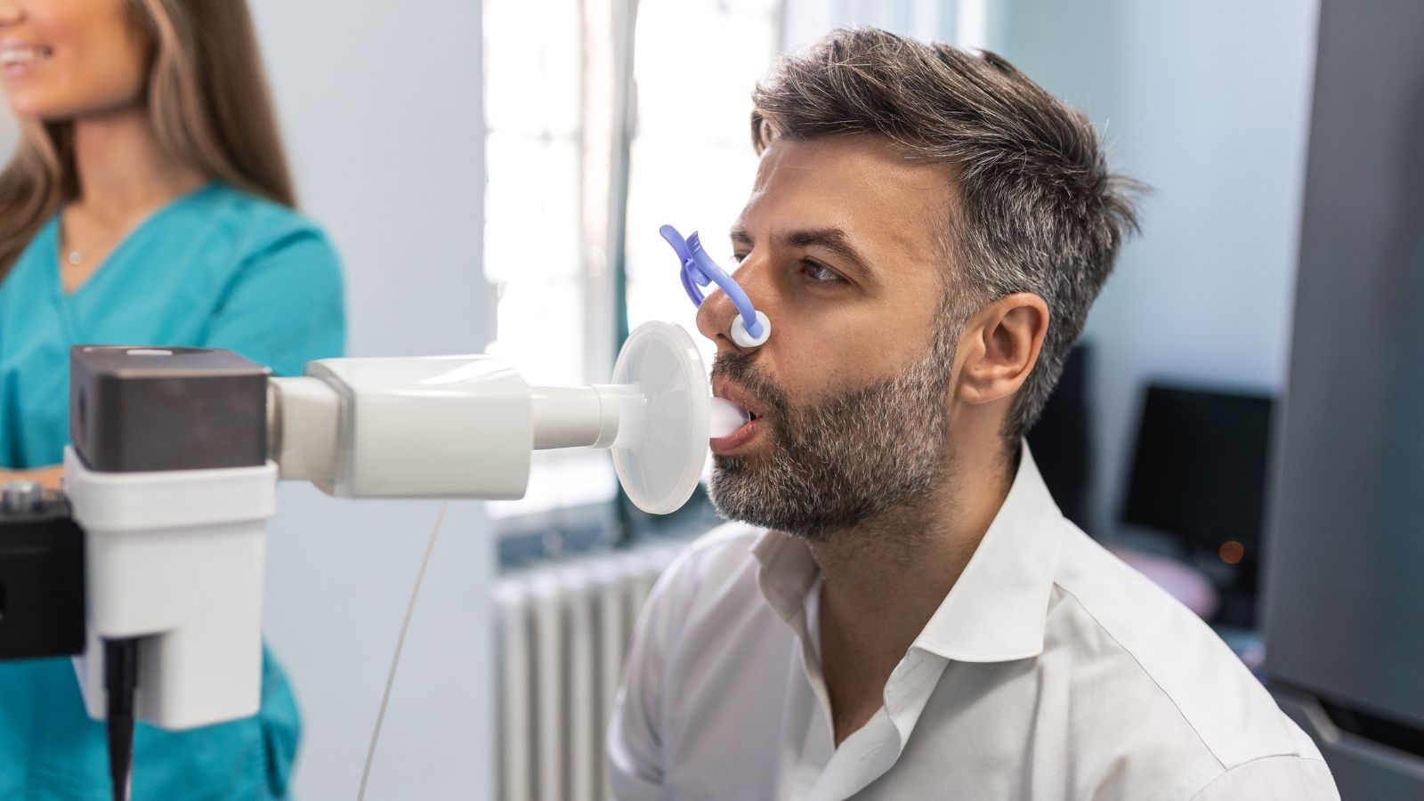
- Pulmonary function tests
- Reporting allergies
- Chest X-ray evaluation
Is Pain Felt During a Lung Biopsy?
Many people mistakenly believe that biopsies are extremely painful procedures. Thanks to modern anesthesia techniques, these concerns are generally unfounded. During needle biopsies, the skin and underlying tissues are completely numbed with powerful local anesthetics similar to those used by dentists. As a result, patients usually feel only mild pressure rather than pain. During minimally invasive surgical biopsies or mediastinoscopy, patients are placed under general anesthesia and remain asleep throughout the procedure, experiencing no pain or awareness.
What Should Be Done During Recovery After a Lung Biopsy?
Patients are not discharged immediately after the procedure. Following needle biopsies, they are generally observed for several hours, undergo follow-up chest X-rays to exclude hidden air leaks or bleeding, and are usually discharged the same day. Following surgical biopsies, patients typically remain hospitalized for one or two nights depending on their condition. During recovery at home, following specific recommendations is essential for restoring full lung function.
Patients who undergo general anesthesia or surgery are especially encouraged to perform deep breathing exercises to reopen small air sacs that may have collapsed. Simple breathing devices containing lightweight plastic balls are commonly used to facilitate these exercises, which significantly accelerate recovery.
Important steps after returning home include:
- Physical rest
- Breathing exercises
- Wound care
- Balanced nutrition
- Medication adherence
- Follow-up appointments
What Are the Possible Risks of a Lung Biopsy?
Like every medical procedure, interventions involving the chest cavity carry certain risks because they involve vital organs within a confined space. However, experienced medical teams are well equipped to anticipate and manage these complications. One of the most common complications is pneumothorax, or lung collapse. If the needle or surgical instrument punctures the lung lining, air may leak into the chest cavity. Small air leaks often resolve on their own with oxygen therapy and observation. Larger air leaks causing breathing difficulty require placement of a chest tube between the ribs to remove the trapped air. Once the lung re-expands, the tube is removed.
Bleeding is another potential complication. Minor injury to small blood vessels may cause patients to notice small streaks of blood in their sputum for one or two days after the procedure. This usually represents only minor bleeding and resolves spontaneously through the body’s normal clotting mechanisms without requiring additional treatment. Under experienced medical supervision, these risks are generally well managed.
When Are Lung Biopsy Results Available and What Do They Tell Us?
Immediately after the biopsy, the tissue samples are placed into special preservative solutions and sent to the pathology laboratory. There, they undergo multiple processing steps before being cut into extremely thin sections and examined under high-powered microscopes by specialist pathologists. Standard pathological analysis generally takes between three and seven days depending on the laboratory’s workload. The initial report determines whether the lesion represents inflammation, a benign tumor, or cancer.
Modern medicine does not stop there. If the lesion proves malignant, additional molecular and genetic analyses are performed on the same tissue sample to identify specific mutations within the cancer cells. These advanced tests may require two to three additional weeks. However, the results allow physicians to prescribe highly targeted therapies or immunotherapy tailored to the tumor’s genetic profile rather than relying solely on conventional chemotherapy.
Frequently Asked Questions

Prof. Dr. Alper Fındıkcıoğlu graduated from Istanbul University Faculty of Medicine and completed his thoracic surgery residency at Dr. Siyami Ersek Hospital. He began his academic career at Başkent University in 2005; he received the title of associate professor in 2014 and professor in 2022. He has extensive experience in lung cancer, chest wall diseases, and minimally invasive surgery. Since 2024, he has been continuing his professional work at Acıbadem Hospital.